Saturday, May 3, 2025

Third degree heart block

Vignette says a 60 year old male presents to the emergency department with complaints of fatigue, dizziness, lightheadedness and occasional near syncopal episodes over the past few days; He also complains of intermittent chest pain and shortness of breath on exertion since the past few days; He has a history of hypertension and takes losartan daily; On examination JVP shows cannon a waves; Vital signs show pulse rate of 35 beats/min which is slow and regular in nature, blood pressure of 110/70 mm of Hg, respiratory rate of 16 breaths/min, oxygen saturation of 96% in RA and temperature of 97.2 F; ECG shows wide QRS complex with no relation between P waves and QRS complexes however R-R intervals and P-P intervals are constant; Diagnosis?


Diagnosis is Third degree heart block.


Third degree heart block is characterized by a complete loss of communication between atria and ventricles (i.e. no impulse from atria passes through the AV node to the ventricles); JVP shows cannon A waves; ECG shows a wide QRS complex (ventricular escape rhythm) or narrow QRS complex (junctional escape rhythm) with no relation between P waves and QRS complexes however R-R intervals & P-P intervals are constant.


Causes:-

1. Idiopathic fibrosis or degeneration of the conducting system (i.e. Lev's disease)

2. Autoimmune disorders (e.g. SLE, systemic sclerosis)

3. Inflammatory conditions (e.g. myocarditis, Lyme disease, acute rheumatic fever)

4. Infiltrative myocardial disease (e.g. hemochromatosis, sarcoidosis, amyloidosis)

5. Electrolyte imbalance (e.g. hyperkalemia)

6. AV nodal blocking agents (e.g. beta-blockers, non-dihydropyridine calcium channel blockers, digitalis, adenosine, or amiodarone)

7. Anterior wall MI (due to extensive necrosis of the septum and conduction tissue i.e. His-Purkinje system)

8. Inferior wall MI (since right coronary artery supplies the AV node (i.e. right coronary dominant))


Presents with generalized fatigue, tiredness, lightheadedness, dizziness, chest pain, shortness of breath, near syncope and syncope. 3rd degree heart block is associated with polyuria (AV dissociation occurs as there is complete loss of communication between atria and ventricles in 3rd degree heart loss; this causes huge stretch in atrium leading to release of ANP which subsequently causes diuresis and polyuria).


Diagnosis:- 

1. ECG shows a wide QRS complex (ventricular escape rhythm) or narrow QRS complex (junctional escape rhythm) with no relation between P waves and QRS complexes however R-R intervals & P-P intervals are constant.

2. Electrophysiology study.


Treatment:-

1. Temporary pacemakers (i.e. transcutaneous or transvenous pacing to increase the ventricular rate). 

2. Insertion of permanent pacemaker (PPM).

3. Treat the underlying cause and stop the offending agents.


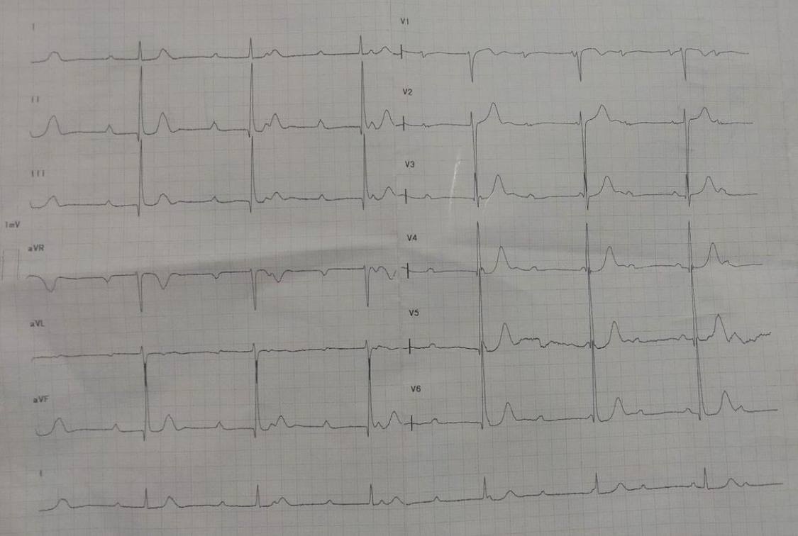
Figure:- ECG showing 3rd degree heart block

Cardiac tamponade

Vignette says a 38 year old male with a history of stage 5 chronic kidney disease on maintenance dialysis presents to the emergency department with chief complaints of progressive shortness of breath, chest discomfort and fatigue over the past few days; He has a history of hypertension, diabetes mellitus and chronic kidney disease and takes medications for hypertension, diabetes and dyslipidemia; Vital signs show blood pressure of 90/60 mm of Hg, pulse rate of 120 bpm, respiratory rate of 22 breaths/min, oxygen saturation of 92% in RA and temperature of 97.2 F; His pulses are thready, weak and becomes even fainter during inspiration; Examination shows jugular venous distension, muffled heart sounds and cool extremities; ECG shows electrical alternans, low voltage QRS complexes; Echocardiography shows pericardial effusion with right ventricular collapse during diastole; Diagnosis?


Diagnosis is Cardiac tamponade.


Cardiac tamponade is a medical emergency condition characterized by excessive accumulation of fluids in the pericardial space restricting the filling of cardiac chambers and subsequent low cardiac output and shock.


Etiologies:-

1. Trauma (penetrating > blunt) 

2. Malignancy (e.g. lung, breast, lymphoma) 

3. Uremia (ESRD) 

4. Pericarditis 

5. Post-MI (especially ventricular wall rupture, Dressler's syndrome) 

6. Iatrogenic (e.g. catheter placement, cardiac surgery)


Pathophysiology:- Increased pericardial pressure → restricts ventricular filling → ↓ preload → ↓ stroke volume → ↓ CO → shock.


Clinical features:-

1. Beck’s triad:- Muffled heart sounds, jugular venous distension and hypotension.

2. Reflex tachycardia, cool extremities, tachypnea with clear lungs.

3. Pulsus paradoxus (i.e. decreased SBP >10 mmHg during inspiration).


Diagnosis:-

1. ECG shows electrical alternans, low voltage QRS complexes.

2. Chest x-ray shows enlarged cardiac silhouette (i.e. water bottle sign).

3. Echocardiography shows pericardial effusion, diastolic collapse of the right atrium and right ventricle, plethoric inferior vena cava (i.e. lack of inspiratory collapse) and impaired ventricular filling.

4. Cardiac catheterization shows elevation and equalization of intra pericardial and diastolic pressures in all chambers of the heart; shows elevated JVP with loss of the x descent.


Treatment:- Immediate pericardiocentesis.

Prinzmetal angina

Vignette says a 20 year old female presents to the cardiology clinic for evaluation of chest pain; She is experiencing increased episodes of central chest pain which is non exertional, self-limiting in nature that typically lasts for brief period (5-15 minutes); The pain is sudden in onset, substernal, tight, and crushing in nature, severe in intensity with radiation to the left arm, jaw and neck associated with nausea, vomiting and diaphoresis; She was diagnosed with Raynaud disease 1 year back but is not under any medications; She doesn't smoke cigarettes but drinks alcohol occasionally; Vital signs are normal; ECG shows ST-segment elevation in leads II, III and aVF during ergonovine test; Coronary catheterization shows coronary vasospasm with no atherosclerotic occlusions; Diagnosis?

Diagnosis is Prinzmetal angina (i.e. variant angina).


Prinzmetal angina occurs due to transient coronary artery vasospasm, leading to reduced blood flow and ischemia in the heart muscle and is characterized by chest pain at rest that is responsive to nitrates with ECG showing transient ST-segment elevation present during chest pain. 

It is characterized by the triad of:- 

1. Chest pain at rest, typically self-limiting and responsive to nitrates. 

2. ECG showing transient ST-segment elevation during chest pain.

3. Coronary catheterization showing coronary vasospasm with no or minimal atherosclerotic occlusions.


Diagnosis:-

1. ECG shows ST-segment elevation during pain episodes.

2. Cardiac biomarkers (e.g. CK-MB, Troponins) are negative.

3. Coronary angiography shows provocative coronary vasospasm (i.e. ergonovine, acetylcholine) with no or minimal atherosclerotic occlusions.


Management:- 

1. Stop the offending agents (e.g. smoking, cocaine, marijuana, triptans).

2. Medical therapy includes nitrates, and CCB's (e.g. verapamil, diltiazem, amlodipine). 

Beta blockers (i.e. nonselective) are contraindicated as they worsen vasospasm.